
2D Gels: Where Protein Patterns Tell the Story
In protein science, not every answer is found in numbers or charts. Sometimes, it’s the visual patterns—the subtle shifts in...
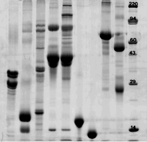
In protein science, not every answer is found in numbers or charts. Sometimes, it’s the visual patterns—the subtle shifts in position, intensity, and form—that reveal what’s really going on in your samples. That’s where 2D gels step in, offering a visual language that proteins speak fluently, if you know how to listen.
As someone invested in understanding proteins—whether for therapeutic development, diagnostics, or food science—you can’t afford to overlook this powerful technique. 2D Gel Electrophoresis transforms your protein data into a map. Every spot, smear, and shape has something to say. And when you’re trained to read it, you uncover biological stories that other tools simply gloss over.
Why 2D Gels Still Matter in a High-Tech Lab
It’s easy to get swept away by mass spectrometry or ultra-fast LC systems. But protein separation isn’t just about speed—it’s about depth, resolution, and context. 2D gels remain the gold standard for:
- Detecting post-translational modifications
- Visualizing protein isoforms
- Comparing complex protein mixtures
- Identifying changes in expression under varying conditions
This method combines Isoelectric Focusing (IEF) with SDS PAGE, giving you a two-dimensional look at your proteome. The result? Proteins are arranged by isoelectric point (pI) in one direction and molecular weight in the other. Patterns emerge, and with them, insights you’d miss in a single-dimension approach.
Reading the Story in Protein Spots
Every gel tells a story. And every protein spot can hold the key to understanding cellular responses, stress markers, disease progression, or bioprocess consistency.
Let’s say you’re working on therapeutic protein development. Subtle changes in pH, temperature, or host cell expression can lead to isoforms or fragments. Without 2D gels, these can go unnoticed. But when laid out across a gel, those variants appear like fingerprints—unique, revealing, and often critical to your end product.
Even in a basic Milk Testing Laboratory, 2D gels can distinguish between casein variants and detect adulterants. In diagnostics, they reveal autoantibodies and disease markers long before symptoms manifest.
Click this Protein Analysis Lab to explore how specialists apply 2D gels across biotech, pharma, and food industries.
What to Watch for in Your Own Gels
Running a 2D gel isn’t just plug-and-play. It requires discipline, technique, and a trained eye. As someone performing or reviewing gel-based work, here’s what you should focus on:
Spot Clarity
Smearing and streaking can obscure results. Clean spots mean clean prep. Use protease inhibitors and keep your samples cold.
Consistency Across Replicates
One gel tells you something. Three matching gels tell you the truth. Watch for consistency to validate any claims about expression changes.
Unexpected Spots
These might be degradation products, contaminants, or modifications. In quality control, they may indicate a failing batch or unapproved process deviation.
Missing Proteins
If key proteins disappear between conditions or treatments, your experiment may have uncovered a regulatory trigger or breakdown.
The more gels you run, the more fluent you become in spotting the significant from the background noise.
Designing Experiments that Speak Clearly
To get a story worth reading from your 2D gels, your experimental setup needs structure:
- Choose pH gradients strategically—narrow gradients like pH 4–7 give more resolution than wide ranges.
- Use matched controls and replicates. Subtle changes are hard to trust without tight comparisons.
- Load equal protein concentrations—too much leads to streaking, too little to faint spots.
In post-run analysis, use digital image software. It’ll let you match spots, calculate densities, and detect expression differences more accurately than the human eye.
From Gels to Insights: What Comes Next
Running the gel is just chapter one. What happens next is where you extract meaning:
Staining
Silver stain is more sensitive than Coomassie. Fluorescent dyes, though more costly, offer multiplexing and digital detection.
Spot Picking
Excise and digest the protein spots for Mass Spectrometry identification. You can confirm post-translational modifications or unknown proteins.
Western Blotting
Confirm the identity of key proteins or isoforms. It’s a good follow-up for critical targets in disease studies or QA settings.
HCP Analysis
In biologics, residual host cell proteins (HCPs) are a serious concern. 2D gels paired with antibody-based detection can reveal unexpected contaminants.
Applications that Go Beyond Research
The beauty of 2D gels is that they’re relevant in both discovery and commercial production:
- In Biopharma, they validate batch consistency and detect unwanted product variants.
- In Academia, they map proteomic changes under stress, mutation, or disease.
- In Food Testing, they verify protein integrity, authenticity, and allergen presence.
- In Environmental Biology, they track microbial responses to pollutants or climate changes.
Wherever proteins are being produced, regulated, or understood, 2D gels offer a visual truth-check.
Avoiding Common Mistakes
If you’re overseeing or running 2D gels, these are some pitfalls to dodge:
- Skipping sample clean-up: Salts and lipids wreak havoc on IEF.
- Using improper pH gradients: If your proteins fall outside the gradient range, you’ll miss them entirely.
- Inconsistent running conditions: Voltage, temp, and buffer changes ruin reproducibility.
- Poor storage: Gels degrade quickly—scan and analyze immediately or store them cold and dry.
When to Call in the Experts
Not every lab has the resources or time to perfect 2D gels in-house. That’s okay. Outsourcing doesn’t mean compromising—if anything, it ensures expertise.
Ask the lab:
- Do they offer Western Blot Gel Electrophoresis post-run?
- Can they quantify spot densities across samples?
- Do they integrate HCP Antibody Coverage with gel workflows?
- Are gels documented digitally for data traceability?
This is your protein story—you want a narrator who knows how to listen and document it properly.
Final Thoughts: Let the Gels Speak
There’s something timeless about 2D gels. In an age of digital noise and automated assays, these gels provide a tangible, visual record of biological truth.
Whether you’re looking at stressed cell lines, validating drug purity, or analyzing food proteins, don’t ignore what 2D gels reveal. They tell the story others miss.
So next time you’re reviewing a protein separation method, ask yourself—what’s being hidden in the single-dimension view? Because with 2D gels, the pattern is the message.
Learn more about applying 2D gels to your workflow and make protein patterns work for you.